
Medial Epicanthoplasty
Medial epicanthoplasty is a minor surgical procedure that corrects the skin fold covering the inner corner of the eye (epicanthal fold) using small Z-plasty flaps or modified techniques. This exposes more of the inner pink corner of the eye (lacrimal caruncle) and elongates the palpebral fissure by approximately 2–4 mm on each side. It is indicated for prominent epicanthal folds and a wide intercanthal distance; it is not recommended when the eyes are already closely spaced.
Compiled from BonBoz Clinic clinical documentation — currently in medical review
Mechanism
An epicanthal fold is a skin fold extending from the upper eyelid that covers the lacrimal caruncle at the inner canthus, making the palpebral fissure appear shorter. Medial epicanthoplasty creates small skin flaps using a Z-plasty or its variations (Park, Hiraga, etc.) to redirect skin tension lines and transpose the flaps. This uncovers the lacrimal caruncle and extends the eyelid margin medially. Little skin is excised; the procedure mainly relies on tissue rearrangement, making outcomes dependent on flap design and skin tension.
Limitations to Know in Advance
Epicanthoplasty can only extend the eye horizontally as far as the lacrimal caruncle and underlying anatomy permit—typically 2–4 mm per side. It cannot truly "bring the eyes closer together" because the bony orbit remains unchanged. If the intercanthal distance is already narrow (equal to or less than the horizontal length of one palpebral fissure), performing an epicanthoplasty can make the eyes look too close together and appear harsh. Over-resection that excessively exposes the pink lacrimal caruncle looks unnatural and is virtually irreversible.
Indications
Epicanthal folds covering part or most of the lacrimal caruncle; an intercanthal distance distinctly wider than the palpebral fissure length; epicanthal folds pulling the double eyelid crease into a hidden or tapered crease at the inner corner. This procedure is commonly combined with blepharoplasty to ensure a smooth, harmonious crease at the inner corner.
Risks to Consider
Scars at the inner canthus stay pink longer than eyelid scars (3–6 months, and potentially longer in thicker skin), scar contracture creating new webbed folds, excessive caruncle exposure, asymmetry, temporary epiphora (watery eyes) due to swelling, and rarely, lacrimal duct injury. For these reasons, the surgeon performs a conservative opening and simulates the results beforehand.
At BonBoz
Our plastic surgeons measure palpebral fissure length, intercanthal distance, and caruncle coverage on standardized clinical photographs. We only recommend epicanthoplasty when facial proportions allow it and will advise against it if it is not suitable. The flap technique is tailored to each individual's skin tension lines. The consulting physician performs the surgery directly, provides inner corner scar care instructions (sun protection, scar gel), and follows up with you for up to 6 months.
Technique variants
1
Mild medial epicanthoplasty
Slightly refine the epicanthal fold while preserving the natural eye shape.
2
Medial epicanthoplasty with crease design
Combined with double eyelid surgery for a seamless crease.
3
Revision medial epicanthoplasty
Correct scar contracture or over-gapping from a previous procedure.
When should you see a doctor?
- Mild epicanthal folds with optimal intercanthal distance — medial epicanthoplasty may not be necessary.
- Thin skin at the inner canthus with a prone tendency to keloids.
- Prior epicanthoplasty resulting in active contracture scars.
- Desire to elongate the eyes beyond anatomical limitations — realistic boundaries must be thoroughly understood before proceeding.
How is it performed?
Local anesthesia is administered. The surgeon marks the flap design (Z-plasty or its variants) at the inner corner of the eye along the direction of the epicanthal fold, makes an incision along the markings, dissects, and transposes the skin flaps to redistribute skin tension. This exposes more of the lacrimal caruncle and extends the eyelid margin medially to the measured extent, followed by closure with ultra-fine sutures. The procedure takes 30–60 minutes for both eyes and is commonly performed in conjunction with blepharoplasty.
Before the procedure
Measure the palpebral fissure, intercanthal distance, and degree of lacrimal caruncle coverage; capture standardized clinical photographs; simulate the projected opening; and discontinue anticoagulants, alcohol, and smoking as instructed.
After the procedure
**Post-Operative Care Instructions:** * **Compress Therapy:** Apply cold compresses during the first 48 hours (days 1–2) for 10–15 minutes per session, spaced 1 hour apart. Switch to warm compresses starting on day 3. * **Hygiene & Medication:** Keep the surgical area clean, cleanse your eyes with sterile saline, and take all medications as prescribed. * **Suture Removal & Follow-Up:** Suture removal takes place on day 7 at the clinic. Scheduled follow-up visits are on day 7, month 1, month 3, and month 6. * **Scar Management:** Pink scars may persist for 1–3 months and will gradually fade by month 6. Use sun protection and apply scar gel as directed. * **Activities & Dietary Restrictions (Avoid for 1 month):** * Do not consume seafood, beef, glutinous rice, water spinach, alcohol, or stimulants. * Do not wear makeup, do not wear contact lenses, and do not rub your eyes. **When to Contact Your Doctor Immediately:** Seek immediate medical attention if you experience: * Severe, abnormal swelling * Pus discharge or heavy bleeding * Fever above 38.5°C * Spreading redness and inflammation * Inability to close your eyes fully after 1 week
Possible risks — your doctor explains them before treatment
- Canthal scars remain pink longer than eyelid scars (3–6 months)
- Scar contracture forming new folds
- Excessive exposure of the lacrimal caruncle
- Asymmetry
- Temporary tearing
- Lacrimal duct injury (rare)
Who should NOT have this?
- Naturally narrow intercanthal distance
- Minimal epicanthal fold, with small eyes primarily caused by drooping eyelids (eyelid crease creation alone is sufficient)
- A pronounced tendency for keloid or hypertrophic scarring
- Active periocular infection or dacryocystitis
- Unrealistic expectations of lengthening the eyes by multiple millimeters or altering the underlying bone structure
How BonBoz uses it
Lower Eyelid Blepharoplasty
Lower eyelid blepharoplasty addresses loose, wrinkled under-eye skin caused by aging through an incision made just below the lower lash line, with optional orbital fat correction. Ideal for individuals where excess skin is the primary concern, typically aged 45–70.
Learn about this method →Full Incisional Double Eyelid Surgery (With Skin and Fat Removal)
Full incisional double eyelid surgery utilizes an incision along the full length of the eyelid crease to remove excess skin, manage fat, and create a stable crease. It serves as a foundational technique for aging upper eyelids and puffy eyelids—not a 'bright-eyed' enlargement procedure.
Learn about this method →Partial Incision Double Eyelid Surgery (Mini Line)
Mini Line double eyelid surgery uses an incision approximately two-thirds the length of the eyelid to remove moderate skin laxity and fix the crease. Positioned between non-incisional techniques and full incisional surgery, it offers greater durability than non-incisional methods while being less invasive than a full incision.
Learn about this method →Eye Procedures: Medial Epicanthoplasty (BonBoz InnerEyes)
Services page: A specialist physician conducts an examination, recommends a suitable treatment plan, and provides transparent pricing before you make your decision.
Want a cost range for your own case?
Estimate the cost →Cost guidance
The cost depends on the extent of correction required, whether it is performed alongside double eyelid surgery, and whether it is a primary or revision procedure.
The final price follows the doctor’s recommendation after an in-person exam. A confirmed quote has no hidden extras.
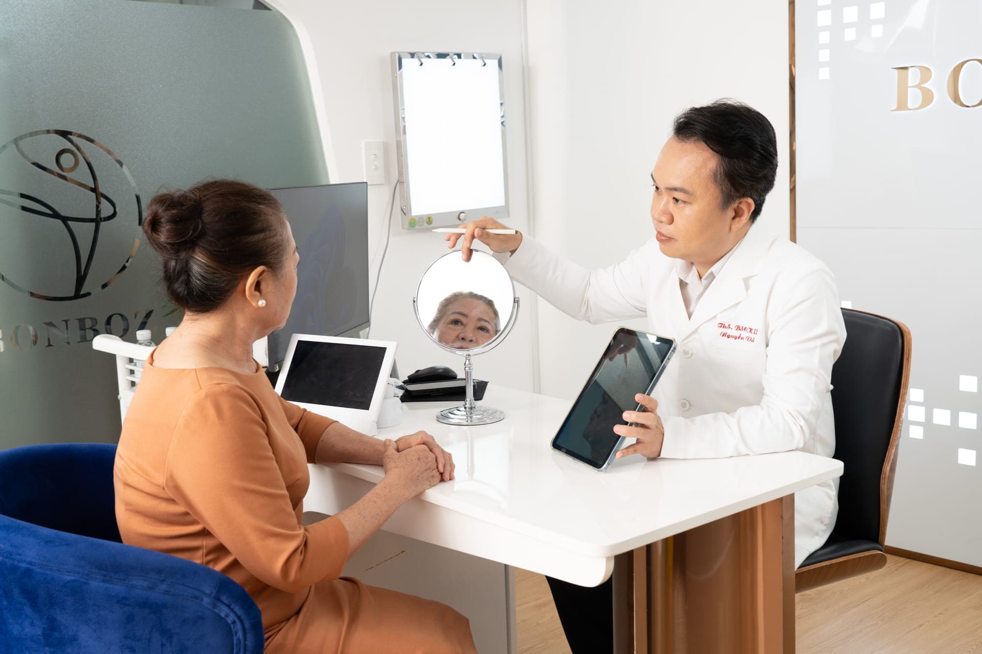
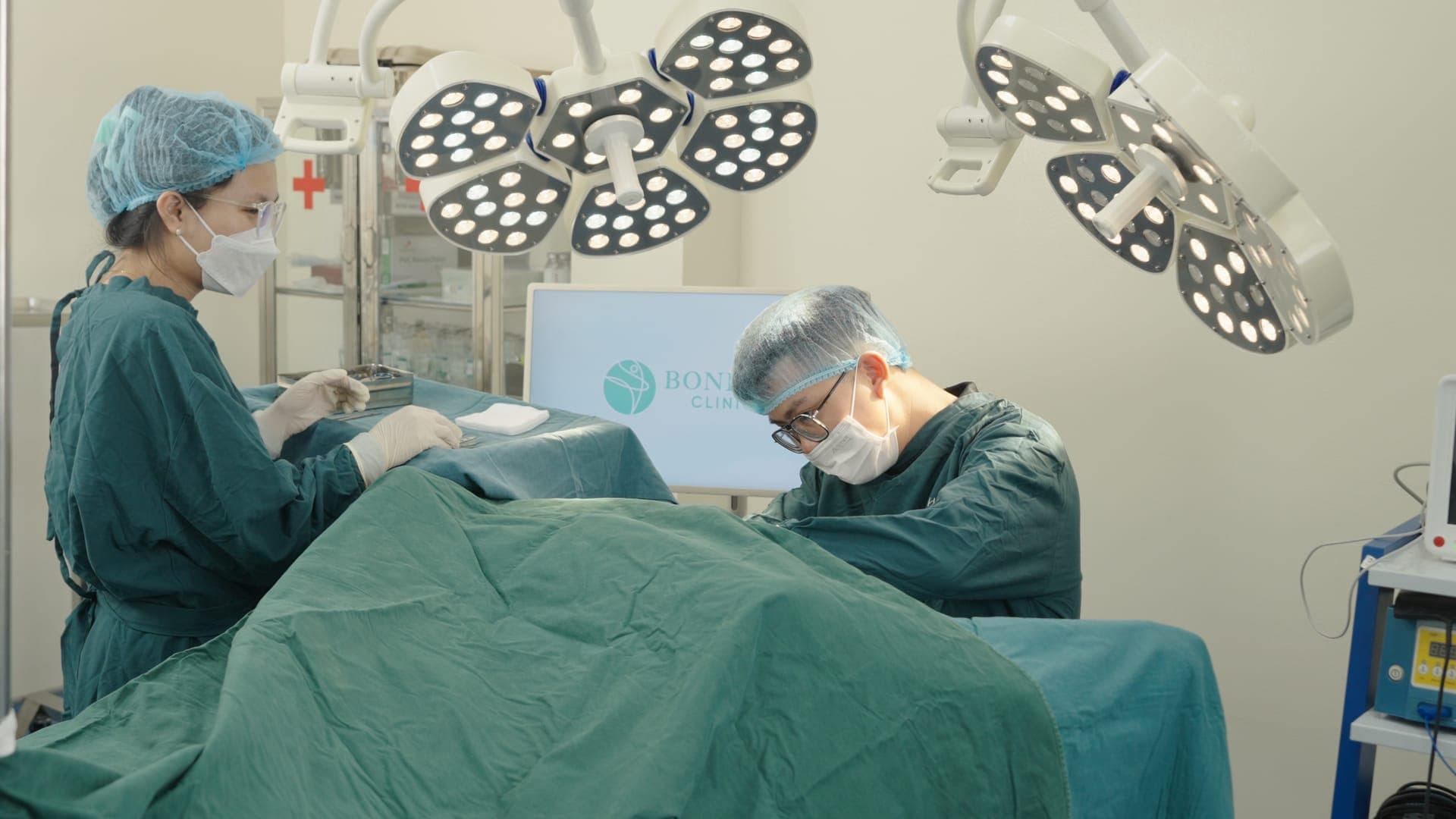
Inside the BonBoz clinic

Book a consultation with a doctor
Choose a date, time slot and doctor — the clinic calls to confirm within 5 minutes during opening hours. Free consultation, no commitment.
Frequently asked questions
How much can medial epicanthoplasty lengthen the eyes?
Are the inner corner scars noticeable?
Can it be performed alongside double eyelid surgery?
Can epicanthoplasty be reversed?
Does it affect the tear ducts?
How long is the recovery time?
If my eyes are close together, should I undergo this procedure?
Can lateral epicanthoplasty be performed at the same time?
Related topics
- Full-Incision Blepharoplasty for Excess Skin and Fat Removal: Procedure, Indications for Aging Eyelids, and Essential Risks to Know
- Mini-Incision Blepharoplasty (Mini Line): Technique, Ideal Candidates, and How It Differs from Full-Incision Blepharoplasty
- Small, Short Eyes with Epicanthic Folds: When to Perform Epicanthoplasty vs. When Double Eyelid Surgery Alone Is Enough
- Monolids vs. Hidden Double Eyelids: What’s the Difference, and Who Should Choose Non-Incisional vs. Incisional Double Eyelid Surgery?
More from our doctors
What are medial and lateral epicanthoplasty? How do they differ, and who is an ideal candidate?
Medial epicanthoplasty and lateral canthoplasty target two distinct anatomical areas of the eye. Understanding what each procedure entails, how they differ, and who is an ideal candidate will help you communicate effectively and precisely with your surgeon.
What Is Double Eyelid Surgery with Epicanthoplasty? How It Differs from Traditional Double Eyelid Surgery
Double eyelid surgery with epicanthoplasty is a surgical technique designed to widen the corners of the eyes, creating larger and more open-looking eyes. This differs from standard upper blepharoplasty, which solely addresses the upper eyelid.
Does Upper Blepharoplasty Actually Make Your Eyes Bigger or Is It Just an Illusion? A Detailed Breakdown
Double eyelid surgery does not actually enlarge the eyes, but it makes them appear brighter and more alert by elevating the eyelid crease and improving facial proportions.